Monday - Friday: 9:00 - 19:00
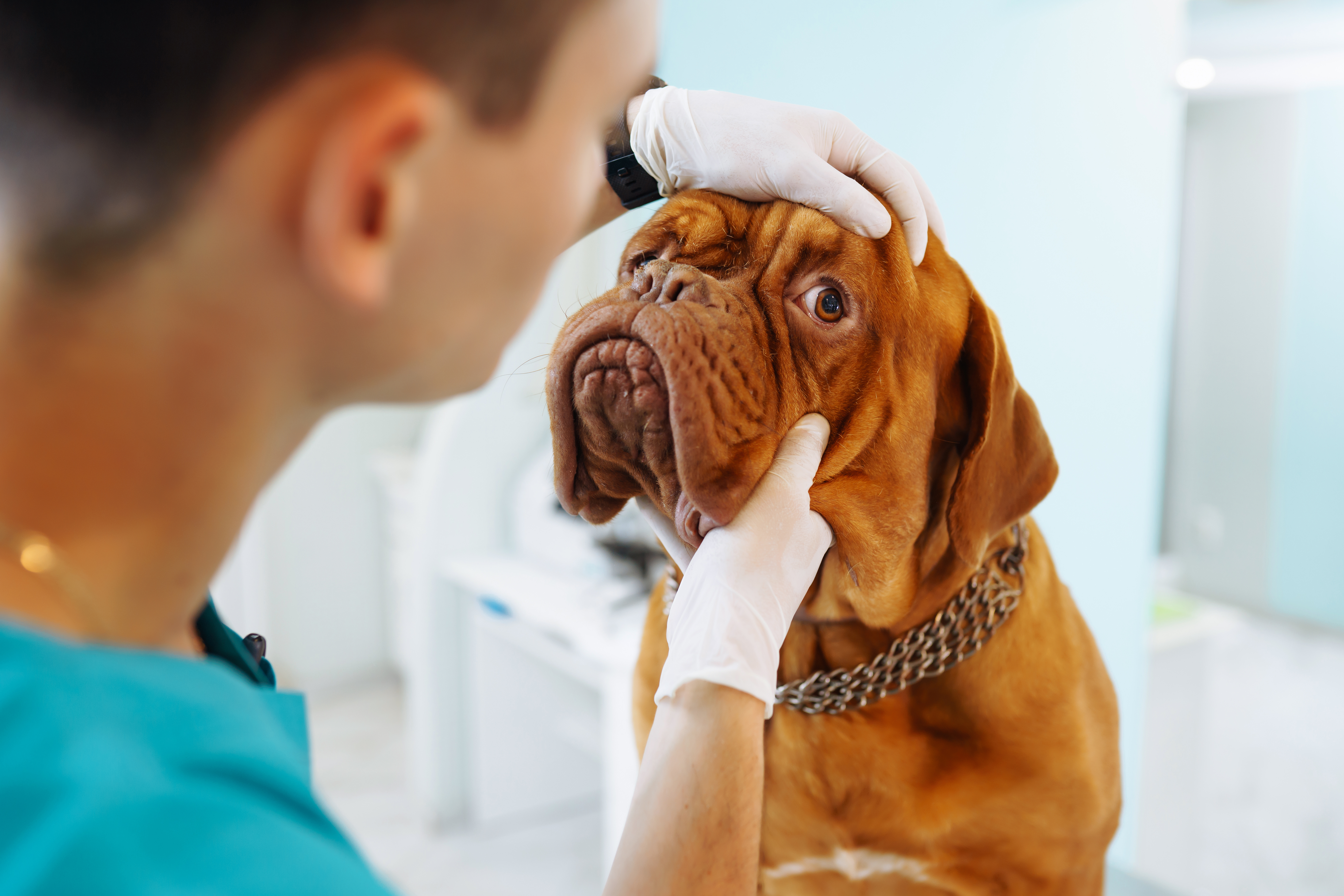
Electroretinography is an eye exam that evaluates the retina's function by measuring tiny electrical impulses generated when light reaches it. This is done using specialised electrodes in the form of a contact lens. Electroretinography can help diagnose various causes of eye problems by detecting retinal lesions early on.
For examination, eye drops are instilled to dilate the pupils and make the surface of the eye insensitive to touch.
During the exam, eye drops dilate the pupils and make the eye surface less sensitive to touch.

To avoid any disruptions in the electrical signals from the retina, the patient is under sedation. A special contact lens with an electrode is then put on the eye. The eye is exposed to bright stimuli of varying intensity and frequency. The electrode records the electrical nerve impulses from the retina that respond to stimuli. This recorded data is then displayed as a computer graph called an "electroretinogram".
Electroretinography is commonly used to detect retinal diseases in their early stages and therapeutic control of retinal diseases and before each cataract surgery.
The risks and complications of anaesthesia are similar to those that can occur during any surgery. Besides that, this examination is painless and without risks. The eyes are light-sensitive for a few hours due to eye drops.
Our team is dedicated to providing you and your pets with the best care possible. Our modern facilities include a laboratory, ultrasound, surgical block with monitoring and gas anaesthesia, and day hospitalisation area.
Our staff values transparency, information, and compassionate care at Madeleine Vet. If you have any questions or need more information, please don't hesitate to ask.

©2023 Madeleine Vet, All rights reserved. Site created with love by Boca